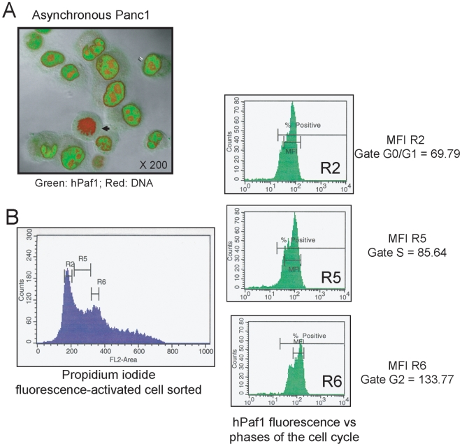
Figure 1. hPaf1 is differentially expressed during the cell cycle.
A) The sub-cellular localization of hPaf1 was determined by confocal analysis of a population of asynchronously growing Panc1 cells that were labeled with FITC-conjugated anti-hPaf1 antibody (green) and counterstained with propidium iodide (red). hPaf1 was detected in the nucleus of the cells, but was absent from the heterochromatin and the nucleoli. hPaf1 was not detected in cells going through mitosis (black arrow). B). The FITC-labeled hPaf1 protein was quantified for each phase of the cell cycle. The mean of fluorescence detected was of 69.79, 85.64, and 133.77 for the cells in the G1-, S- and G2-phases, respectively.

