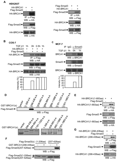
Figure 3. BRCA1 and Smad3 association.
(A) HA-BRCA1 interacts with Flag-Smad3 in HEK293T cells. 0.5 µg of wild type HA-BRCA1 and 0.50 µg of Flag-Smad3 plasmids were co-transfected into HEK293T cells, as indicated. 36 hrs later, total cell lysates were subjected to immunoprecipitation and Western blotting with anti-Flag monoclonal antibody and anti-HA-monoclonal antibody, as indicated. 10 µg of total cell lysates was used to examine the expression levels of Flag-Smad3 and HA-BRCA1 protein. The blots are representative experiment out of 3 experiments. (B) TGF-β1 increases the interaction between HA-BRCA1 and Flag-Smad3 in COS-7 cells. 0.50 µg of Flag-Smad3 and 0.50 µg of HA-BRCA1 plasmids were co-transfected into COS-7 cells. 24 hrs later, cells were treated with or without 2 ng/ml of TGF-β1 as indicated. Total cell lysates were immunoprecipitated with anti-Flag monoclonal antibody followed by Western blotting with anti-HA monoclonal antibody. The membrane was re-probed with anti-Flag antibody. The increases in HA-BRCA1 were normalized to Flag-Smad3 and are represented graphically. The expression levels of HA-BRCA1 protein were detected in 10 µg of total cell lysates. This is a representative experiment out of 3 experiments. (C) TGF-β1 increases BRCA1 and Smad3 interaction in MCF-7 cells. Cells were stimulated with 2 ng/ml of TGF-β1 for 1 hour. Total cell lysates were immunoprecipitated by anti-Smad3 polyclonal antibody, followed by Western blotting with anti-BRCA1 antibody. The membrane was re-probed with anti-Smad3 antibody. The increases in BRCA1 were normalized to Smad3 and are represented graphically. The expression levels of BRCA1 protein were determined in 30 µg of total cell lysates. This is a representative experiment out of 4 experiments. (D) Smad3 binding site in BRCA1. 10 µg of the individual GST-BRCA1 protein fragments and HEK293T cell lysates transfected with Flag-Smad3 plasmid were subjected to a GST-pull down assay. To monitor expression levels, 10 µg of individual GST-BRCA1 protein fragments were separated on 15% and 8% SDS-PAGE, followed by Coomassie staining. (E) Interaction of BRCA1 (1–683aa) with Smad3 protein in HEK293T cells. 0.5 µg of HA-BRCA1(1–683aa) and 0.5 µg of Smad3 plasmids were co-transfected into HEK293T cells, followed by immunoprecipitation and Western blotting, as indicated. The membrane was re-probed with anti-Flag monoclonal antibody to examine the levels of Smad3 protein expression. The levels of HA-BRCA1(1–683aa) expression were determined in 10 µg of total lysates. (F) BRCA1 binds to the MH2 domain in Smad3. 10 µg of GST and GST-BRCA1 (298–436aa) proteins and HEK293T cell lysates transfected with Flag-Smad3 (1–206aa) or Flag-Smad3 (207–426aa) were subjected to a GST pull-down assay. The expression levels of Flag-Smad3 (1–206aa) and Flag-Smad3 (207–426aa) protein were determined in 10 µg of total cell lysates. This is a representative experiment out of 4 experiments. (G) Flag-Smad3 and HA-BRCA1 (298–436aa) interaction. HA-BRCA1 (298–436aa) and Flag-Smad3 plasmids were co-transfected into HEK293T cells, followed by immunoprecipitation and Western blotting, as indicated. The membrane was re-probed with anti-Flag antibody to monitor Flag-Smad3 expression. 10 µg of total cell lysates was used to examine the expression levels of HA-BRCA1 (298–436aa) protein.

