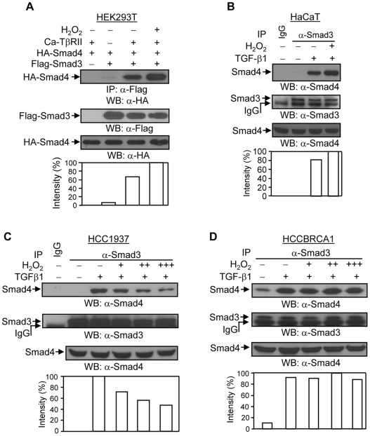
Figure 5. Effects of H2O2 on the TGF-β1-induced association between Smad3 and Smad4.
(A) H2O2 increases the interaction between Smad3 and Smad4 in HEK293T cells. HEK293T cells were co-transfected with 0.5 µg of Flag-Smad3, 0.5 µg of HA-Smad4, and 0.15 µg of caTβRII plasmids, as indicated. 24 hours later, cells were treated with 200 µM of H2O2 for 2 hours, followed by immunoprecipitation and Western blotting, as indicated. The membrane was re-probed with anti-Flag monoclonal antibody to examine the expression levels of Flag-Smad3 protein. The increases in Flag-Smad3-binding to HA-Smad4 were normalized to Flag-Smad3 protein expression and are represented graphically. The expression levels of HA-Smad4 protein were determined in 10 µg of total cell lysates. This is a representative experiment out of 4 experiments. (B) H2O2 increases the interaction between Smad3 and Smad4 in HaCaT cells. HaCaT cells were treated or untreated with 2 ng/ml of TGF-β1 and 100 µM of H2O2 for 1 hour as indicated, and subjected to immunoprecipitation with anti-Smad3 polyclonal antibody and Western blotting with anti-Smad4 monoclonal antibody. The membrane was re-probed with anti-Smad3 antibody to examine the levels of Smad3 protein. The increases in Smad3-binding to Smad4 were normalized to Smad3 protein expression and are represented graphically. The expression levels of Smad4 protein were determined in 20 µg of total cell lysates. This is a representative experiment out of 4 experiments. (C) TGF-β1-induced Smad3 and Smad4 interaction is decreased by H2O2 in HCC1937 cells. HCC1937 cells were treated or untreated with 2 ng/ml of TGF-β1 and 100 (+), 200 (++), and 300 µM (+++) of H2O2 for 1 hour as indicated. Immunoprecipitation was done as in Figure 5b (HaCaT cells). The membrane was re-probed with anti-Smad3 antibody to examine the levels of Smad3 protein. The decreases in Smad3-binding to Smad4 were normalized to Smad3 protein expression and are represented graphically. The expression levels of Smad4 protein were determined in 20 µg of total cell lysates. This is a representative experiment out of 4 experiments. (D) Wild type BRCA1 restores the TGF-β1-induced Smad3 and Smad4 interaction against H2O2 in HCCBRCA1 cells. The experiment was done as in Figure 5c (HCC1937 cells). The increases in Smad3-binding to Smad4 were normalized to Smad3 protein expression and are represented graphically. This is a representative experiment out of 4 experiments.

