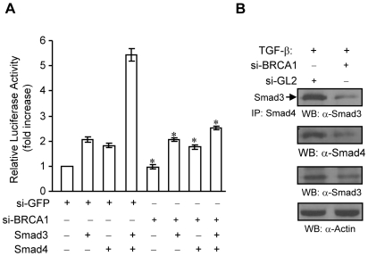
Figure 7. Effects of silencing BRCA1 in MCF-7 cells on Smad3 and Smad4.
(A) si-BRCA1 decreases Smad3-Smad4-mediated transcriptional activation of p3TP-Lux reporter in MCF7 cells. MCF7 cells were transfected with 5 nM of siRNAs in 24 well plate. After 16–24 hours, the cells were transfected with p3TP-Lux, pCMV-β-galactosidase, Smad3, Smad4, and empty vector plasmids as indicated. The cells were maintained in cell culture medium containing 0.1% FBS overnight, followed by luciferase and β-galactosidase assays. The fold increase of luciferase activity was obtained from triplicate of three independent experiments, after being normalized by β-glactosidase activity. The Excel software was used to calculate the standard deviation. * P<0.05 as compared to si-GFP treatments+Smad3+Smad4. (B) Depletion of BRCA1 decreases TGF-β-induced Smad3-Smad4 interaction in MCF-7 cells. MCF-7 cells were transfected with 5 nM siGL2 or siBRCA1. After overnight, cells were starved for 3 hours, followed by 1 ng/ml of TGF-β1 treatment for 45 min. Total lysates were subjected to immunoprecipitation with anti-Smad4 antibody and Western blotting with anti-Smad3 antibody as indicated. The membrane was re-probed with anti-Smad4 antibody to monitor the levels of Smad4 protein. The levels of Smad3 protein was detected from 20 µg of total lysatesby using anti-Smad3 antibody. This membrane was reprobed with anti-Actin antibody to monitor equal loading.

