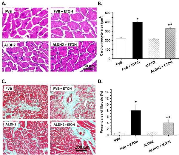
Fig. 4.
Histological analyses hearts from FVB and ALDH2 mice with or without chronic alcohol intake for 14 weeks. A: Representative H&E staining micrographs showing transverse sections of left ventricular myocardium (x 400); B: Quantitative analysis of cardiomyocyte cross-sectional (transverse) area using measurements of ∼ 200 cardiomyocytes from 3-5 mice per group; C: Representative Masson trichrome staining micrographs showing longitudinal sections of left ventricular myocardium (x 200); and D: Quantitative analysis of fibrotic area (Masson trichrome stained area in light blue color normalized to the total myocardial area). Data were obtained from 3-5 mice per group, *p < 0.05 vs. FVB, # p < 0.05 vs. FVB+ETOH.

