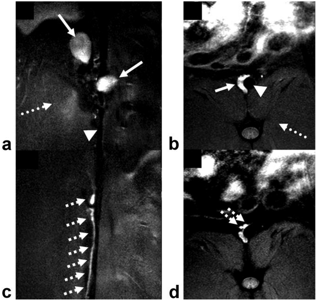
Figure 3.
Postcontrast coronal (a,c) and axial (b,d) fast spin echo images using IRON demonstrate high contrast enhancement in paraaortic lymph nodes (solid arrows, a,b) and in the paraaortic lymphatic ducts (hatched arrows, c,d), while the abdominal aorta (arrowheads, a,b) and the adjacent muscles are signal attenuated (dotted arrows, a,b).

