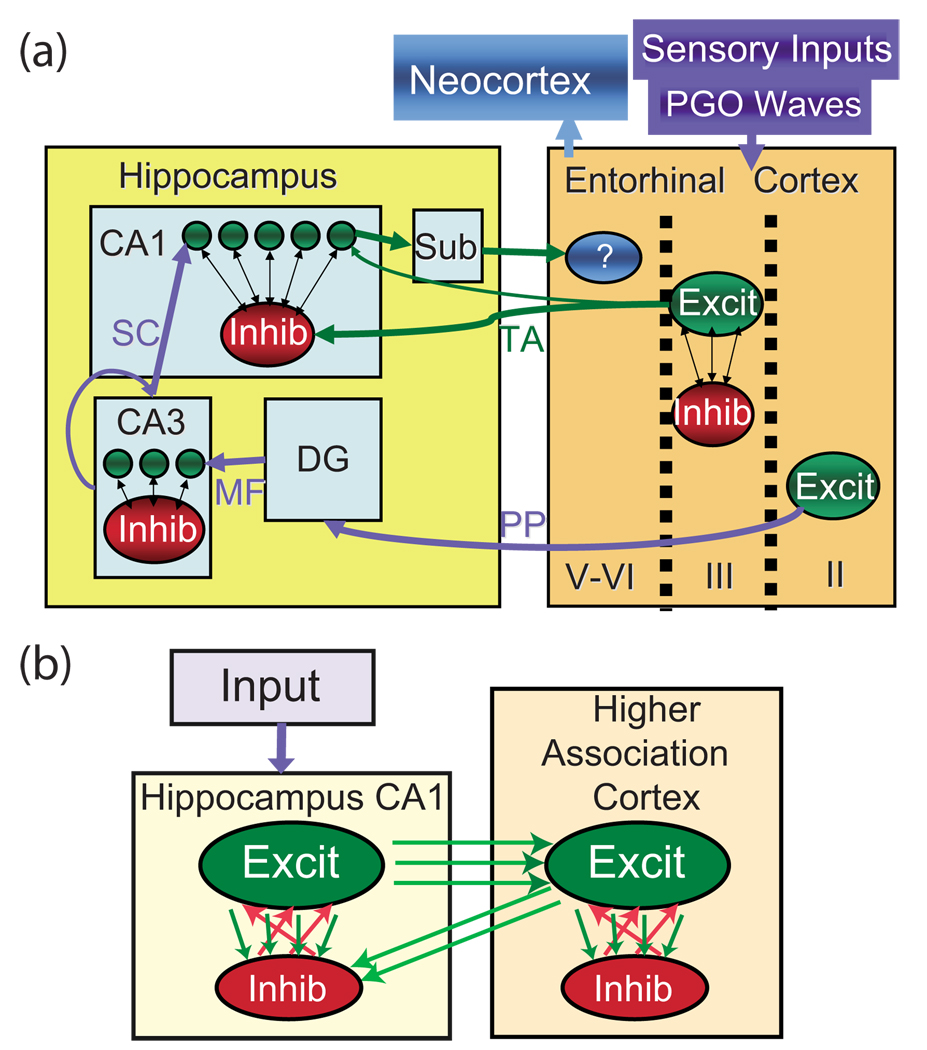
FIG. 1.
(Color online) Diagrams of network structure. (a) Circuit diagram of anatomical connectivity between hippocampal and cortical structures. Entorhinal cortex layers II, III, and IV–VI project through the perforant path (PP) to the dentate gyrus (DG) and CA3, through the temporoammonic (TA) path to the subiculum (sub) and CA1, and from the CA1 and sub to the deeper layers of the entorhinal cortex, respectively. MF=mossy fibers and SC = shaffer’s collaterals. (b) Diagram of model used in simulations. Single network (hippocampus or cortex): the network is composed of a larger population of excitatory neurons and a smaller population of inhibitory neurons. Both inhibitory and excitatory networks are small-world networks having periodic boundary conditions. Feedback between hippocampus and cortex: the excitatory hippocampal neurons locally innervate the excitatory cortical network (e.g., the entorhinal cortex). The cortical excitatory network suppresses the hippocampal excitatory network through random inhibitory pathways.

