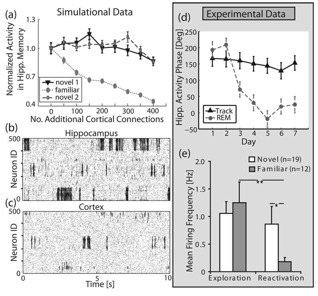
FIG. 4.
Selective autonomous memory reactivation in the hippocampal-cortical structure—simulations and experimental data. The hippocampus has three regions of network inhomogeneities imbedded (IDs 1–100, 200–300, 400–500). One memory structure (IDs 200–300) is stored in the corresponding region in the cortex. (a) Average reactivation activity of familiar (neuron IDs 200–300) vs. novel (neuron IDs 1–100 and 400–500) memories as a function of additional cortical connections. The hippocampal heterogeneity, which has progressively stronger representation in the cortex, reactivates significantly less. Activity was measured by normalizing total spike counts within a memory region for the total duration of the run to total spike counts for the homogenous cortex run. Sample raster plots for (b) hippocampus and (c) cortex. (d), (e) Experimental data: (d) Phase locking of hippocampal neurons activity to peak of cortical theta oscillations as a function of days of exposure to the stimulus (i.e., stimulus novelty). Solid black line (“track”) denotes phase locking to CA1 layer theta peaks during active exploration; dashed gray line denotes the same phase relation observed during REM sleep reactivation (“REM”). The neural firing becomes progressively in-phase with peak theta oscillation observed at distal cortical TA inputs. (e) frequency of hippocampal activity in novel and familiar environments during exploration (left) and sleep reactivation (right).

