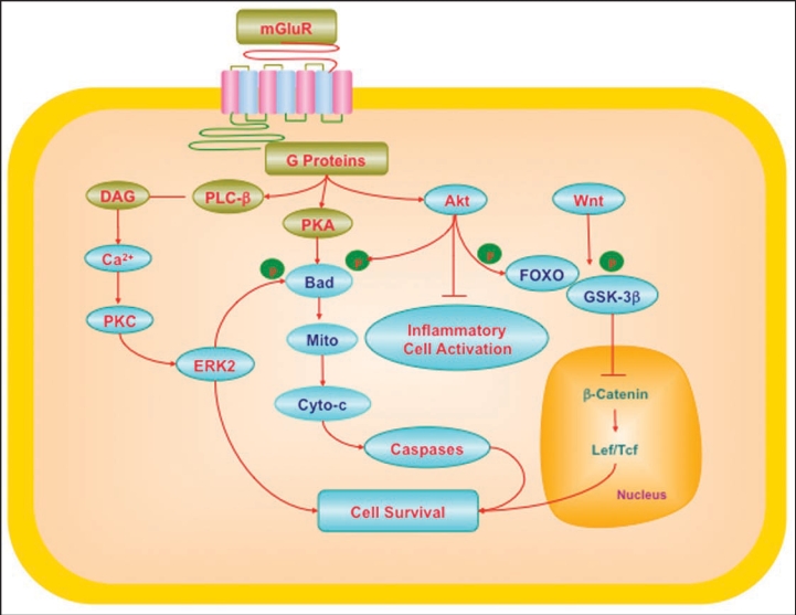
Figure 1.
Cytoprotection through mGluRs may require the regulation of multiple cellular pathways. G-protein βγ in the mGluR system activates phospholipase β (PLC-β), diacylglycerol (DAG), and phosphoinositide 3 kinase (PI 3-K). These pathways lead to the activation of protein kinases A (PKA), B (Akt) and C (PKC) that can involve intracellular calcium (Ca2+). mGluRs can activate ERK2 through PKC. PKA can phosphorylate (p) Bad, a member of Bcl-2 protein family, which can prevent apoptotic cell injury. Akt provides an anti-apoptotic survival signal through the phosphorylation and inactivation of Bad and also interfaces with the Wnt and forkhead pathways. Wnt signaling can inhibit glycogen synthase kinase (GSK-3β). Inhibition of GSK-3β prevents phosphorylation (p) of β-catenin and leads to the accumulation of β-catenin. β-catenin subsequently translocates to the cell nucleus and contributes to the formation of Lef/Tcf lymphocyte enhancer factor/T cell factor (Lef/Tcf) and β-catenin complex that may cooperate with factors to target nuclear gene transcription and prevent cell injury. Interconnected pathways with Akt and Wnt involve the forkhead family members (FOXO), mitochondrial (Mito) membrane potential (ΔΨm), cytochrome c (Cyto-c), and caspases. If left unabated, these pathways converge to lead to apoptotic cell injury.

