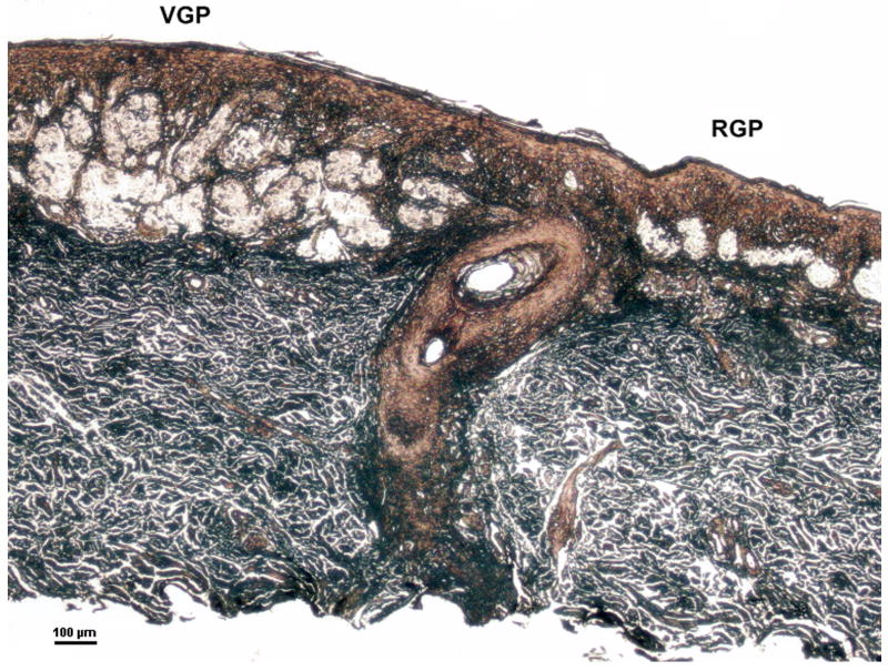
Figure 1.
Microdissection of melanoma cells from the radial and vertical growth phases of a cutaneous melanoma. The sample represents #26 in Table 2. Microdissected tumor cells from the VGP (left) had invaded the dermis whereas RGP tumor cells (right) were localized within the epidermis. Hematoxylin and eosin staining, modified for laser capture microdissection; scale bar = 100 um.

