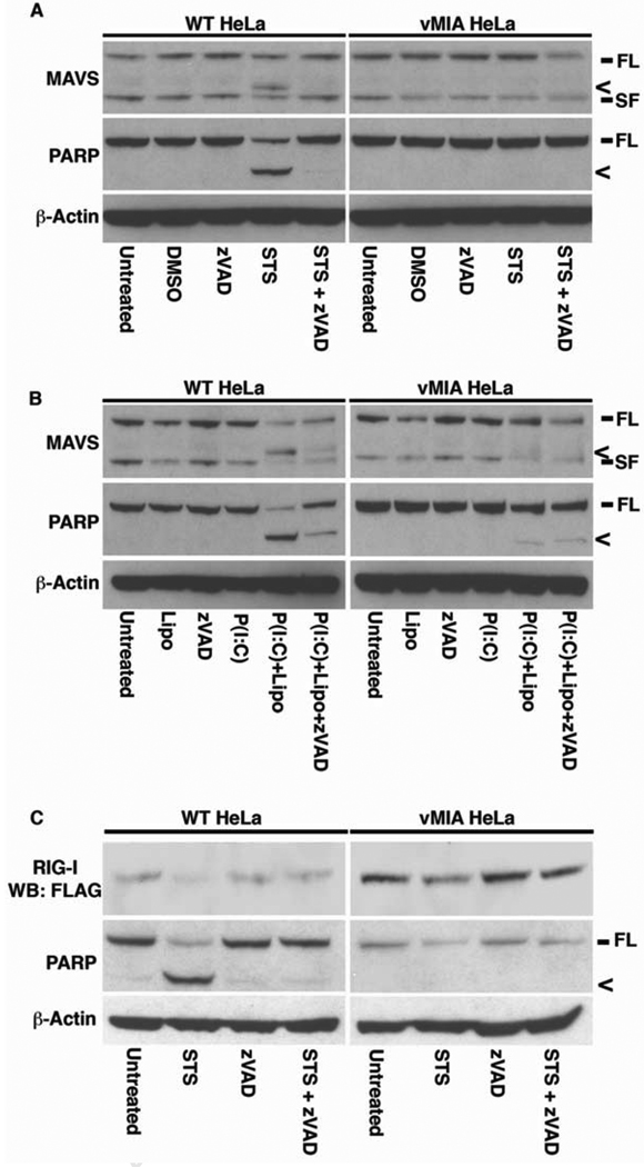
Figure 1. vMIA protects MAVS and RIG-I from capsase-mediated cleavage during apoptosis.
A WT and vMIA HeLa cells were treated with 0.5 µM staurosporine (STS) in the presence or absence of 50 µM zVAD-fmk (zVAD). Untreated and vehicle-only (DMSO) cells were used as controls. After 2 h, cells were harvested and 50 µg of protein extracts were analyzed for MAVS and PARP by western blotting, with β-actin being used as a loading control. B WT and vMIA HeLa cells were treated with 5 µg ml−1 Poly (I:C) for 6 h, either added to the growth medium (P(I:C)), or transfected into cells (P(I:C) + Lipo) using Lipofectamine 2000, in the presence or absence of 50 µM zVAD. 50 µg of protein extracts were analyzed for MAVS, PARP and β-actin as above. C WT and vMIA HeLa cells were transfected for 16 h with FLAG-RIG-I, then treated for 2 h with 0.5 µM STS in the presence or absence of 50 µM zVAD. 50 µg of protein extracts were analyzed for RIG-I (α-FLAG), PARP and β-actin as above. Symbols: FL – full-length protein; SF – short-form of protein, < – cleavage product.

