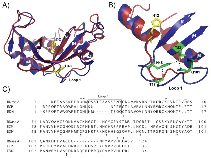
Figure 1.
Structural comparison of bovine ribonuclease A (RNase A) and human Eosinophil Cationic Protein (ECP). A) Superposition of RNase A (blue structure, PDB code 1FS3 (35)) and ECP (red structure, PDB code 1DYT (33)). H12 highlights the position of the active site relative to the position of His48 and loop 1 in RNase A. B) Zoomed view of a few atomic interactions between His48 and loop 1 in RNase A. Note the longer loop 1 in RNase A (D14SSTSAASSSNY25) relative to the shorter loop 1 of ECP (S17LNPPR22, ECP numbering). C) Primary sequence alignment of RNase A, ECP and EDN. Active site positions Gln11, His12, Lys41, His119 and Asp121 are marked with stars while cysteine residues forming the strictly conserved four disulfide bridges are represented by arrows. Loop 1 and position 48 are boxed. Alignment was performed with TCoffee Expresso (36) using PDB coordinates 7RSA (32), 1DYT (37) and 1GQV (38).

