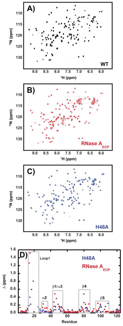
Figure 2.
Chemical shift changes caused by mutation. 1H-15N HSQC spectra for (A) wild-type, (B) RNase AECP, and (C) H48A enzymes. The spectra were acquired at 14.1 T and 298K, pH = 6.4. In (D) chemical shift differences for WT vs. H48A (blue) and WT vs. RNase AECP are shown as a function of amino acid sequence. Gray boxed areas depict protein regions with larger than average chemical shift disturbances. (Δ (ppm) = [(Δδ2HN + Δδ2 N/25)/2]1/2) in which δ is the difference in chemical shift between the two proteins for NH and HN nuclei (52).

