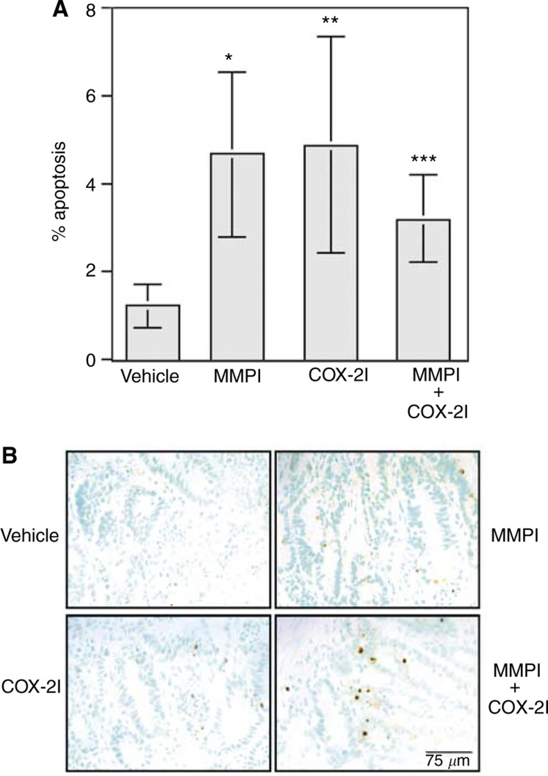
Figure 3.
Tumours from Min mice treated with MMPI or COX-2I have increased levels of apoptosis in comparison with vehicle-treated mice. Intestinal tumour sections were stained for apoptosis using the ApopTag kit and number of positive nuclei and total tumour-associated nuclei were counted as described in Materials and Methods. (A) A total of 1000 nuclei were counted per tumour. Results are represented as percent nuclei staining positive. Each bar is representative of total number of tumours scored for each group where vehicle n=11, MMPI n=10, COX-2I n=10, and combination n=6. Statistical differences were calculated as described in Materials and Methods by comparing vehicle and treated groups. *P=0.0012, **P=0.0018, ***P=0.009. (B) Representative staining for apoptosis from each treatment group. Apoptotic nuclei are stained brown and contrast green is used to visualize total nuclei.

