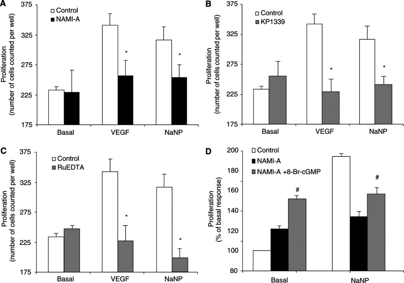
Figure 5.
Proliferation of postcapillary endothelial cells in the presence of VEGF or exogenous NO. (A–C) Cells were incubated in the absence and in the presence of NO scavengers (3 μM NAMI-A, 3 μM KP1339 or 30 μM RuEDTA). Cell proliferation was monitored after 48 h incubation. Data (means±s.e.m.) are reported as number of cells counted per well. (D) Cell proliferation in response to 10 μM NaNP was studied in endothelial cells exposed to 3 μM NAMI-A in the presence of 100 μM 8-Br-cGMP (n=3 in triplicate). *P<0.05 vs control and #P<0.05 vs NAMI-A alone.

