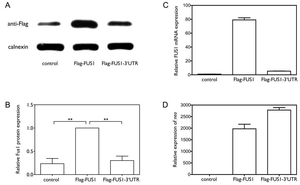
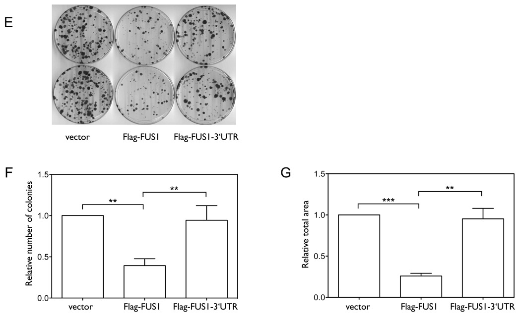
Figure 1. The 3’UTR plays a significant role in regulating Fus1 protein and mRNA expression levels.
(A–D) We cloned constructs expressing either Flag-tagged FUS1 without the 3’UTR (Flag-FUS1) or Flag-tagged FUS1 with the full length 3’UTR (Flag-FUS1-3’UTR). NCI-H1299 cells were transfected for 48 h with either equal amounts (0.46 nM) of the above constructs, or were treated with only transfection reagents (control). (A) The protein expression level of Fus1 was detected by Western blot using an anti-Flag antibody, with calnexin as a loading control. (B) Quantification results of the Fus1 protein level were from three independent experiments. (C) The mRNA level of FUS1 as detected by quantitative PCR. (D) The mRNA level of neo as detected by quantitative PCR. (E–G) Colony formation s a function of the presence or absence of the FUS1 3’UTR. NCI-H1299 cells were transfected with equal amounts (0.46 nM) of either constructs expressing Flag-FUS1, Flag-FUS1-3’UTR or empty vector (control), respectively. Colonies were visualized by staining with 1% crystal violet (E). Quantification of (F) number of colonies and (G) total area of colonies was from three independent colony formation assays. (**, p<0.01; ***, p<0.001)


