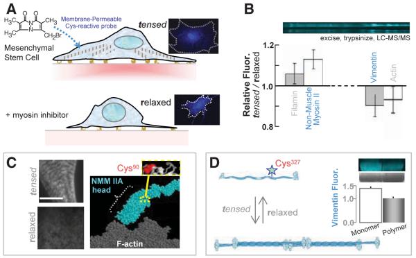
Fig. 4.
In-cell labeling of human stem cells either tensed or relaxed. (A) The membrane-permeable Cys-reactive fluorophore, mBBr, was added to 1-week cultures of MSCs with active myosins and tensed cytoskeletons and also to MSCs treated with myosin-inhibiting blebbistatin for 1 day to relax the cells. Imaging shows homogeneous labeling with 0.5 mM mBBr for 40 min. (B) SDS-PAGE and densitometry of samples (±SD, three experiments) that were either blebbistatin treated (relaxed) or untreated (tensed) show several protein bands in which the fluorescence intensities are different (normalized to protein load). Lysates were quenched with β-mercaptoethanol (50 mM) before analysis. (C) Immunofluorescence imaging of NMM IIA in tensed cells (top) and relaxed cells (bottom) indicates a spatial redistribution of myosin with drug. Scale, 5 μm. MS analyses of excised myosin bands detected labeling of Cys90, which appears buried within the fold of NMM IIA homology models. (D) Vimentin labeling in monomeric, and polymeric forms display different degrees of fluorescence (error bar from two experiments), which indicates that polymerization sterically blocks Cys327 for labeling.

