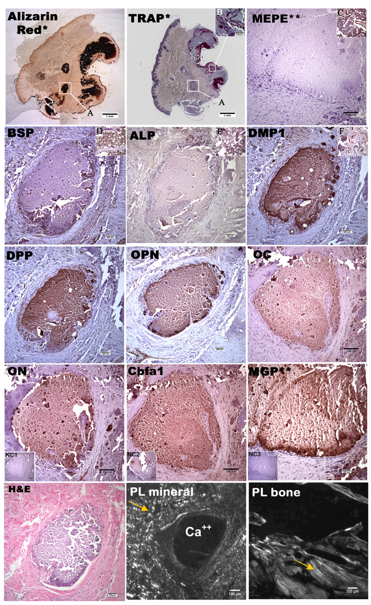
Figure 1.
Alizarin red stain, TRAP stain, H&E stain and immunohistological stain for DPP, OPN, MEPE, BSP, DMP1, ALP, Cbfa1, ON, OC, MGP in mineral deposits and adjacent connective tissue taken at 10x magnification. Brown indicates positive stain. *picture taken at 1x magnification, **picture taken at different site, NC1 is negative control for DPP, OPN, MEPE, BSP, DMP1, ALP, ON, OC,; NC2 is the negative control for Cbfa1; NC3 is the negative control for MGP (A) magnified region in the other panels, (B) indicated region of TRAP positive osteoclasts at 10x magnification (C) human bone as positive control for MEPE, (D) positive control for BSP, (E) positive control for ALP, (F) representative 63x magnified region of surrounding cells demonstrating DMP1 reactivity within the cytoplasm; (PL Mineral) represents JDM mineral deposits under polarized light; (PL bone) represent normal bone taken under polarized light. Arrows indicate collagen.

