Figure 2.
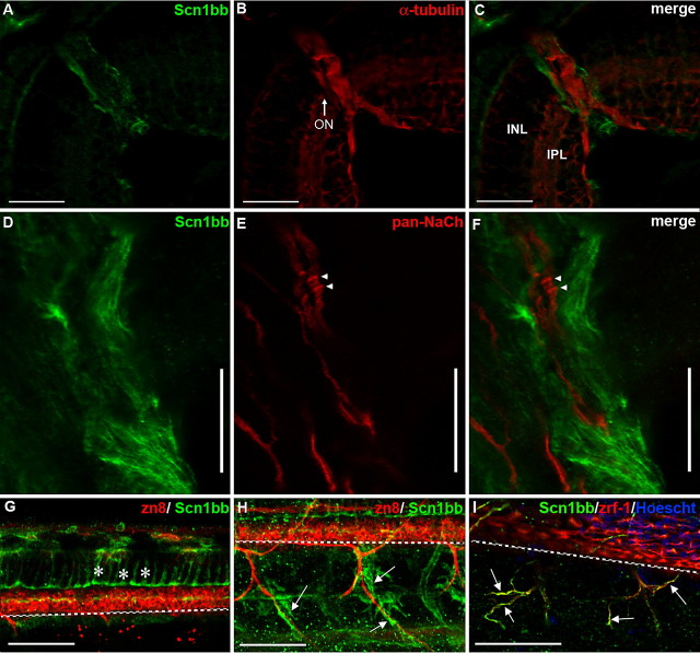
Scn1bb is expressed in optic nerve and spinal cord glia. A–C, Cryosections; scale bars, 25 μm. A, At 5 dpf, the retina displays little or no Scn1bb immunoreactivity (green). However, cells surrounding the optic nerve (ON) are strongly Scn1bb immunopositive. B, Acetylated α-tubulin (red) staining is observed in layers of the retina (e.g., inner plexiform layer, IPL) and strongly labels the optic nerve (ON). C, The merged image of A and B demonstrates that Scn1bb (green) and acetylated α-tubulin (red) immunoreactivites do not overlap in the optic nerve. In addition, the Scn1bb signal surrounds that of the neuronal marker acetylated α-tubulin, indicating that Scn1bb immunopositive cells localize to the location of optic nerve myelin. D–F, Dissected optic nerves; scale bars, 10 μm. D, We detected strong Scn1bb immunoreactivity in optic nerves dissected from adult zebrafish. E, In addition to Scn1bb, isolated adult optic nerves display axonal expression of pan-Na+ channel α-subunits (NaCh, red). Na+ channel α-subunit clusters, possibly hemi-nodes, are indicated with arrowheads. F, The merged image shows that Scn1bb (green) and Na+ channel α-subunit (red) immunoreactivities are nonoverlapping, consistent with Scn1bb expression in the myelinating glia that ensheath the retinal ganglion cell axons within the optic nerve. G–I, Lateral view of whole mount 72 hpf embryos, anterior to the left, dorsal at the top. Scale bars, 50 μm. The dashed white line indicates the ventral boundary of the spinal cord. G, Radial glia (asterisks) in the spinal cord express Scn1bb (green) and can be identified by their characteristic elongated morphology, stretching from the dorsal spinal cord to just above the secondary motor neurons (labeled with zn8, red). H, Scn1bb-positive cells (green, arrows) surround, but do not overlap with, ventral motor neuron axons (labeled with zn8, red). I, Scn1bb (green) colocalizes with the Schwann cell marker zrf-1 (red), suggesting Scn1bb is expressed in Schwann cells (arrows) that ensheath peripheral motor axons.

