Figure 6.
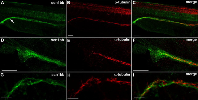
Scn1bb is expressed in the epithelial cells of the pronephric duct. A, Anti-Scn1bb (green) labeled the cells spanning the pronephric duct (arrow) in 28 hpf fish. B, Anti-acetylated α-tubulin (red) labeled the cilia that line the pronephric duct. C, Anti-Scn1bb (green) and anti-acetylated α-tubulin (red) colabeled the pronephric duct. D, Anti-Scn1bb (green) labeled epithelial cells of the pronephric duct. E, Anti-acetylated α-tubulin (red) labeled the ciliated interior of the pronephric duct. F, Expression of scn1bb (green) and acetylated α-tubulin (red) is nonoverlapping. G, High magnification image of anti-Scn1bb positive (green) epithelial cells. H, High magnification image of anti-acetylated α-tubulin positive (red) cilia. I, Merged image with expression of Scn1bb (green) and acetylated α-tubulin (red) nonoverlapping. Scale bars: A–F, 50 μm; G–I, 10 μm.

