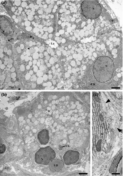
Electron micrographs of control (a) and diabetic (b, c) SMG acinar cells. Control acinar cells show basally located nuclei and rough ER; the apical secretory granules have a light, flocculent content and occasionally fuse with their neighbours. Cells of diabetic animals contain basally located lipid droplets (LD) and occasional residual body-type lysosomes (LY). Thickening and redundancy of the basal lamina (arrowheads, panel c) occur in diabetic glands. AC, acinar cell; N, nucleus; Lu, lumen; IC, intercellular canaliculus. a and b, scale bars = 2 μm; C, scale bar = 0.5 μm.

An official website of the United States government
Here's how you know
Official websites use .gov
A
.gov website belongs to an official
government organization in the United States.
Secure .gov websites use HTTPS
A lock (
) or https:// means you've safely
connected to the .gov website. Share sensitive
information only on official, secure websites.
