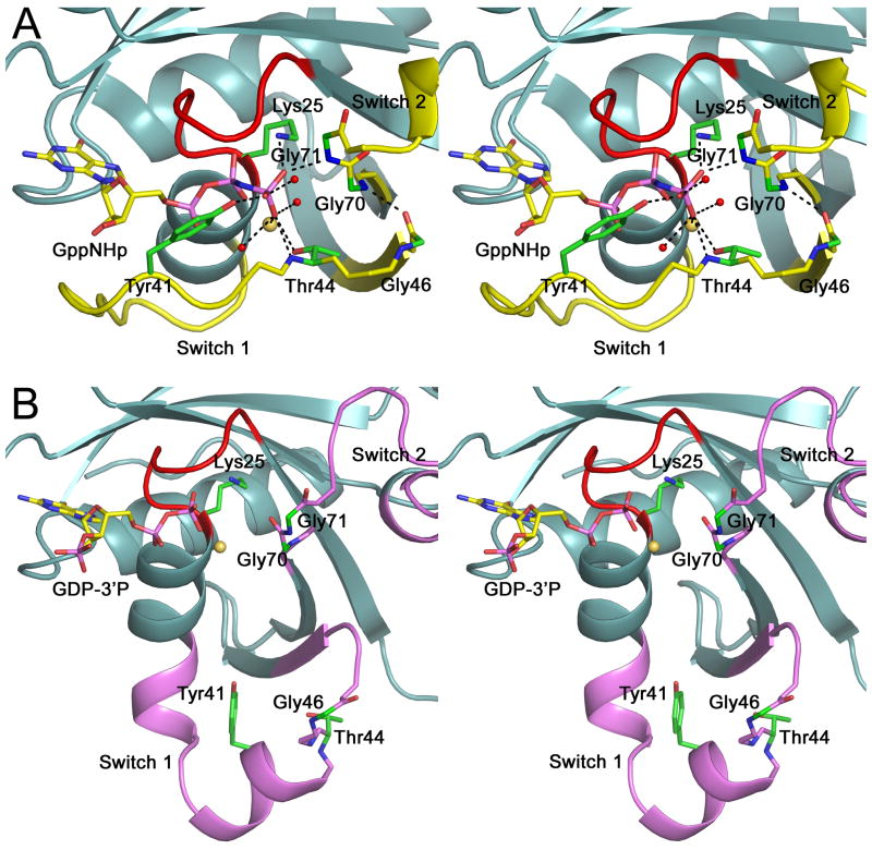
Fig. 3.
The double-glycine motif and the conformational change. Close-up stereo views of the nucleotide, the double-glycine motif and the switch region in the structures of GppNHp-Rab28 (A) and GDP-3′P-Rab28 (B). The hydrogen-bonding network around the γ-phosphate of the nucleotide is broken after hydrolysis and phosphate release, resulting in destabilization of the double-glycine motif and the conformational change in the switch region. Colors are according to figures 1A and 2A.

