Fig. 2.
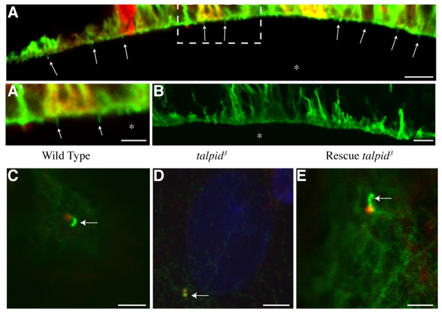
Rescue of primary cilia. (A,B) Rescue of primary cilia in talpid3 mutant neural tube after electroporation with construct encoding full-length chicken Talpid3. (A) Section of electroporated side of neural tube expressing RFP, showing rescue of primary cilia, indicated with arrows, projecting into lumen (*) and stained with acetylated tubulin (green). (A′) Higher magnification of outlined area in A; rescued primary cilia are indicated with arrows. (B) Section of non-electroporated side of neural tube, no primary cilia present. (C-E) Rescue of primary cilium formation in talpid3 mutant CEFs. (C) Wild-type CEF with primary cilium axoneme stained with acetylated tubulin, (green; arrow) protruding from centrosome stained with γ-tubulin (red). (D) talpid3 mutant CEF no primary cilia; acetylated tubulin staining (green) almost entirely overlaps with centrosome staining (red, arrow). (E) talpid3 mutant CEF transfected with a construct encoding full-length chicken Talpid3 shows rescue of primary cilium, arrow indicates axoneme protruding from centrosome. Scale bars: 8 μm in A,B; 4 μm in A′; 3 μm in C-E.

