Fig. 4.
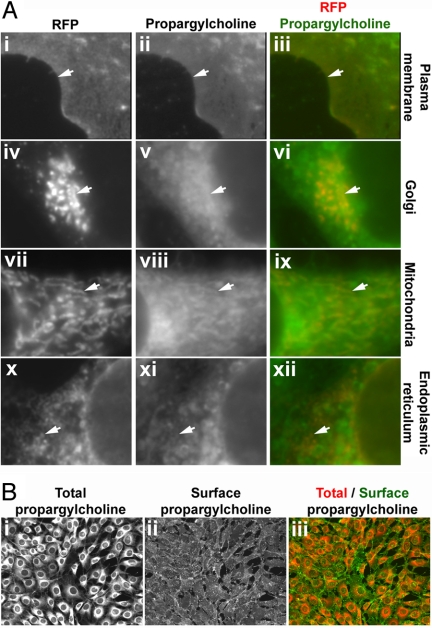
Subcellular distribution of propargylcholine-labeled phospholipids. (A) Co-localization of the propargyl-Cho stain with subcellular markers. Cultured NIH 3T3 cells were transfected with plasmids encoding red fluorescent protein fusions that mark various organelles. The cells were labeled with 100 μM propargyl-Cho overnight and stained with fluorescein-azide. Propargyl-Cho co-localizes with markers for the plasma membrane (i–iii), the Golgi (iv–vi), mitochondria (vii–ix), and endoplasmic reticulum (x–xii). The white arrows point to subcellular structures that stain for both propargyl-Cho and a given red fluorescent marker. (B) Staining propargyl-Cho phospholipids localized to the outer leaflet of the plasma membrane. Cells labeled with 100 μM propargyl-Cho were reacted with Alexa568-azide and biotin-azide. The latter is visualized specifically on the cell surface by staining with Alexa488-conjugated streptavidin (ii), which due to its size does not cross the plasma membrane. The right panel (iii) shows the overlay of the total (i) and surface (ii) propargyl-Cho stain.

