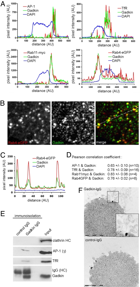
Fig. 2.
Gadkin associates with AP-1- and TfR-containing endosomal organelles. (A–D) Colocalization of endogenous Gadkin with AP-1, TfR, Rab11-myc, and Rab4-eGFP in HeLa cells. (A) Fluorescence intensity profiles of HeLa cells immunostained for endogenous Gadkin and TfR, AP-1, transfected Rab11-myc, or Rab4-eGFP. (B) Enlarged view of the peripheral area of a HeLa cell expressing Rab4-eGFP (depicted in Fig. S1D) immunostained for endogenous Gadkin. (C) Line profile taken at the indicated position in B. (D) Pearson correlation coefficients for the colocalization between Gadkin and AP-1, TfR, Rab11-myc, or Rab4-eGFP. (E) Immunoblot analysis of immunoisolated Gadkin-containing organelles. Shown are representative immunoblots (three independent experiments) using control IgGs or affinity-purified IgGs monospecific for Gadkin (see also Fig. S3G). Input, 50 μg starting material. Samples were analyzed by SDS-PAGE and immunoblotting for CHC, AP-1γ, TfR, and Gadkin. (F) Thin-section electron microscopy of organelles immunoisolated using Gadkin-specific (Top) or control IgG (Bottom) as described in E. The dark material seen in the images represents Dynal (M-280) magnetic beads used for immunoisolation. Inset, 2-fold magnified image of boxed area. (Scale bar, 1 μm.)

