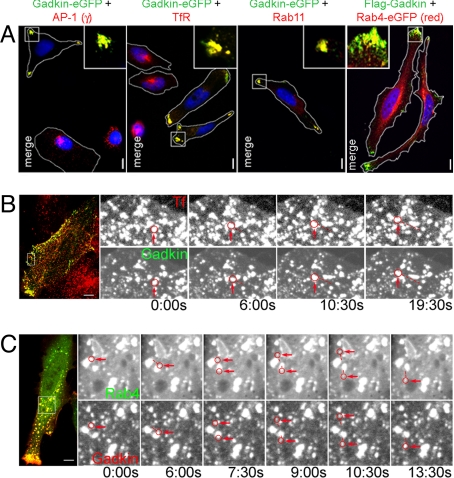
Fig. 3.
Ectopically expressed Gadkin induces the dispersion of EVs. (A) Overexpression of Gadkin-eGFP or Flag-Gadkin causes peripheral dispersion of AP-1- and TfR-containing Rab11- and Rab4-positive EVs. HeLa cells expressing Gadkin-eGFP or Flag-Gadkin (24 h posttransfection) were analyzed by indirect immunofluorescence microscopy with antibodies against AP-1γ, TfR, and Rab11. Rab4 was detected via its eGFP-tag. Cell boundaries are outlined in white. Blue, DAPI-stained nuclei. Insets, 3× magnified views of boxed areas. (Scale bar, 10 μm.) (B and C) A subpool of motile Gadkin vesicles is positive for Tf and Rab4. (B) HeLa cells expressing Gadkin-eGFP loaded with Alexa568-Tf (1 h at 37 °C) analyzed by live-cell confocal imaging at 37 °C. Shown are still images (enlargements of the boxed area of the cell depicted on the Left) extracted from Movie S1 at the indicated time points. (C) Still confocal images (enlargements of the boxed area of the cell depicted on the Left) extracted from Movie S2 of a Gadkin-mRFP and Rab4-eGFP coexpressing HeLa cell. Examples of motile vesicles positive for both markers are circled in red and pointed at. The distances traveled by the vesicles are indicated by dotted lines. (Scale bar, 10 μm.)

