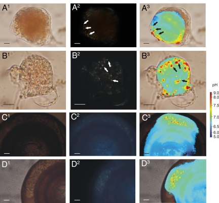
Fig. 4.
Occurrence of calcite needles and correlated high pH vesicles in juvenile specimens of Quinqueloculina yabei and adults of Cyclogyra planorbis. Pictures of all series are taken at the same time. (A and B) 1: Bright field microscopical pictures of juvenile Q. yabei, 2: Polarized light indicating the calcite crystallites inside the cell (white arrows), and 3: Intracellular pH distribution, superimposed on bright field picture (1) containing high pH vesicles correlated to the location of the crystallites (black arrows). (C and D) Same series of pictures for C. planorbis. (Scale bar, 10 μm.)

