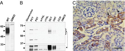
Fig. 1.
Glut9 is expressed in the apical and basolateral membrane of distal convoluted tubules. (A) Western blot analysis of Glut9 expression (arrowhead) in whole kidney lysates from wild type (WT) and G9KO mice. The C-terminal directed antibody recognizes both the Glut9a and b isoforms. (B) Western blot analysis of Glut9 expression (arrowhead) in the indicated nephron segments: PCT, proximal convoluted tubule; PST: proximal straight tubule; mTAL: medullary thick ascending loop of Henle; cTAL: cortical thick ascending loop of Henle; DCT: distal convoluted tubule; CNT: connecting tubule; CCD: cortical collecting duct; OMCD: outer medulla collecting duct. A longer exposure time (lower panel) reveals a marginal expression of Glut9 in the PCT and PST. (C) Immunohistochemical detection of Glut9. Immunoreactivity is evident in the basolateral (black arrows) and apical (white arrowheads) membranes of cells from the distal tubule. (Scale bar, 50 μm.)

