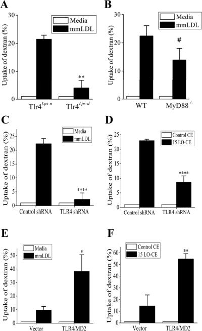
Figure 2. TLR4-dependent uptake of dextran.
A and B, Peritoneal resident macrophages from TLR4-competent and TLR4-deficient C3H mice (A) and from MyD88-knockout and wild type C57BL6 mice (B) were incubated with media alone or 50 μg/ml mmLDL for 1 hour in the presence of Alexa Fluor-488 labeled dextran (10,000 Da). The dextran uptake was measured by FACS and presented as percent change in the geometric mean of FACS histograms. **, p<0.01 (n=4); #, not significant (p=0.150, n=6).
C and D, TLR4-knockdown and control J774 macrophages were incubated with media alone or 50 μg/ml mmLDL for 1 hour (C), or 2.5 μg/ml of non-oxidized CE or 15LO-CE for 15 min (D) in the presence of Alexa Fluor-488 labeled dextran (10,000 Da). ****, p<0.0005 (n=5).
E and F, CHO cell lines expressing human TLR4/MD-2 or empty vector were incubated with media alone or 50 μg/ml mmLDL for 1 hour (E), or 2.5 μg/ml of non-oxidized CE or 15LO-CE for 15 min (F) in the presence of Alexa Fluor-488 labeled dextran (10,000 Da). *, p<0.05; **, p<0.01 (n=5).

