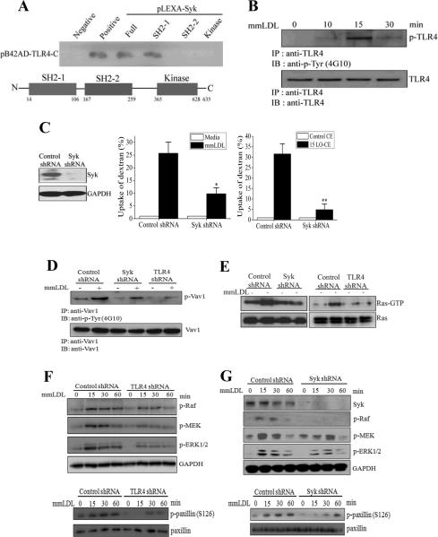
Figure 3. Signaling of mmLDL-induced, TLR4-dependent macropinocytosis.
A, Yeast two-hybrid analysis of binding of the C-terminal domain of TLR4 with the full length Syk, two SH2 domains and the kinase domain of Syk.
B, J774 macrophages were incubated with 50 μg/ml mmLDL for indicated times. Cell lysates were immunoprecipitated with a TLR4 antibody and probed with an antibody against phosphotyrosine (upper panel) or TLR4 (lower panel).
C, Control and Syk-knockdown J774 macrophages (the Syk-knockdown was confirmed in an immunoblot as shown in left-hand panel) were incubated with media alone or 50 μg/ml mmLDL for 1 hour (middle panel), or 2.5 μg/ml of non-oxidized CE or 15LO-CE for 15 min (right-hand panel), in the presence of Alexa Fluor 488-labeled dextran (10,000 Da). The dextran uptake was measured by FACS and presented as an increase in the geometric mean of FACS histograms compared to media or control CE. Mean±standard error from 3 to 5 independent experiments. *, p<0.05; **, p<0.01.
D, Control, Syk-knockdown and TLR4-knockdown J774 macrophages were incubated with 50 μg/ml mmLDL for 30 min. Cell lysates were immunoprecipitated with a Vav1 antibody and probed with an antibody against phospho-tyrosine (upper panel) or Vav1 (lower panel).
E, Control, Syk-knockdown and TLR4-knockdown J774 macrophages were incubated with 50 μg/ml mmLDL for 15 min. Cell lysates were tested for Ras-GTP and total Ras.
F and G, Control, TLR4-knockdown and Syk-knockdown J774 macrophages were incubated with 50 μg/ml mmLDL for indicated times. Cell lysates were probed for phosphorylated Raf, MEK1 and ERK1/2, and GAPDH as a loading control, as well as for phosphorylated and total paxillin. The Syk knockdown was confirmed by probing the lysates with a Syk antibody, and the TLR4 knockdown was confirmed in a FACS assay as in Online Figure I.

