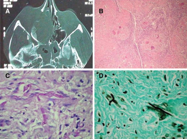
Fig. 2.
(A) Computed tomography scan of patient with chronic granulomatous fungal rhinosinusitis involving the right nasal cavity in a chronic invasive granulomatous fungal rhinosinusitis with bony destruction of paranasal sinuses extending into right orbit. (B) Extensive granulomatous process in a fibrotic background on hematoxylin and eosin stain (×100). (C) Fungal hyphae inside giant cells on periodic acid-Schiff stain (×400). (D) Fungal hyphae inside giant cells on Gomori methenamine-silver stain (×400).

