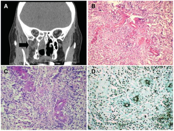
Fig. 3.
(A) Coronal computed tomography scan of immunosuppressed patient with amyloidosis and chronic invasive mucormycosis in chronic invasive fungal rhinosinusitis. Right ethmoid and pterygopalatine space involvement. (B) Nongranulomatous chronic inflammatory infiltrate with transverse section of fungal hyphae eosinophilic Splendore-Hoeppli phenomenon on hematoxylin and eosin stain (×200). (C) Periodic acid-Schiff stain (×400). (D) Gomori methenamine-silver stain (×200).

