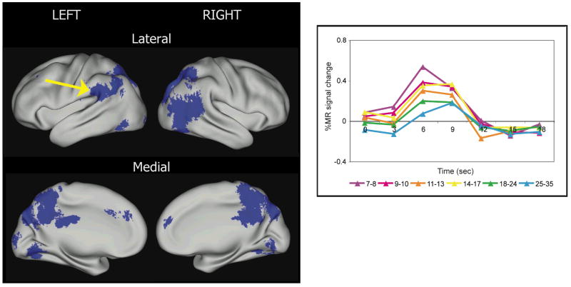
Fig. 2.
Regions that are significantly different over age for high-frequency single word reading and auditory repeating tasks (adapted from Church, Coalson, Lugar, Petersen, & Schlaggar, 2008). All regions decreased in activity over age and are shown in blue. The brain images are organized in the same manner as described in Figure 1. At right, the time courses of blood-oxygen level-dependent signal change for the reading task in six incremental age bins for 81 people are shown on a graph for a left parietal region where activity decreases over age (arrow). This region is in a location hypothesized to be important in phonological processing, and the higher level of activity in children suggests that adults have a relatively decreased reliance on phonological processing when reading aloud high-frequency words.

