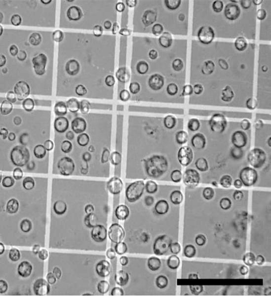
Figure 2. Bright field image of a purified protoplast fraction.
A. parasiticus was grown for 36 h under aflatoxin inducing conditions and protoplasts prepared and purified as described in Methods. 10μL of the pure protoplast fraction were dispensed into a hemacytometer and observed by bright field microscopy to estimate purity and total number. Size bar = 50μm.

