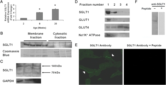
Figure 1.
SGLT1 is expressed in murine and human cardiac myocytes. (A) Relative SGLT1 mRNA expression was assessed by QPCR in hearts harvested from male WT FVB mice at ages 2, 8, and 20 weeks (n = 5 per group). Data are expressed as mean ± SE. *P < 0.01 relative to 2-week-old hearts. (B) A representative immunoblot of membrane and cytosolic fractions of murine cardiac protein showed that SGLT1 was present only in the membrane fraction. Coomassie blue staining of the protein gel was used to document the relative quantity of protein loaded for the immunoblot. (C) A representative immunoblot of total human cardiac protein showed the presence of two (70 and 140 kDa) SGLT1 bands in all lanes, and an intermediate band in the rightmost lane. An immunoblot of GAPDH was used to document the relative quantity of protein loaded. (D) A representative immunoblot of murine cardiac protein fractions derived on a sucrose gradient showed colocalization of SGLT1, GLUT1, and Na+/K+ ATPase (a marker for the sarcolemma). (E) Immunofluorescence microscopy showed that SGLT1 was predominantly localized to the sarcolemma of cardiac myocytes from 8-week-old male WT FVB mice (left, arrowheads), a staining pattern that was significantly decreased by pre-incubation of the antibody with the immunizing peptide. (F) Further to demonstrate anti-murine SGLT1 antibody specificity, the SGLT1 band visualized by immunoblot was completely competed off by pre-incubation of the antibody with the peptide.

