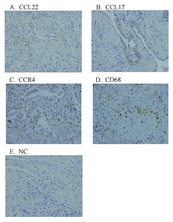
Figure 3.
Lung immunohistochemical photomicrograph of CCL17, CCL22, CCR4, and CD68 in patients with idiopathic pulmonary fibrosis (IPF). We examined the localization of CCL17, CCL22, CCR4, and CD68 by immunohistochemistry. The sections were initially incubated with anti-CCL22 antibody (A), anti-CCL17 antibody (B), anti-CCR4 antibody (C), anti-CD68 antibody (D), or their diluent buffer (E), and then stained using an indirect streptavidin-biotinylated complex method. A fraction of the alveolar macrophages was positive for CCL22, whereas CCL17 was exclusively expressed by some hyperplastic epithelial cells (A, B). There were few alveolar macrophages which were weakly positive for CCR4 (C). The tissue distribution of alveolar macrophages was confirmed by their positivity for CD68 (D). In contrast, no lung cells were positively stained in negative control (NC) sections (E).

