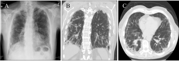
Figure 1.
A. Plain chest radiograph shows bibasilar infiltrates with a peripheral reticulonodular pattern superimposed on generalized interstitial changes involving the upper lobes and lung bases. B. (Coronal view) and C. (Axial View) High Resolution Computed Tomography of the chest shows thickening of intralobular septa, septal line formation, parenchymal band formation and peribronchial thickening. Mild mediastinal lymphadenopathy is also noted.

