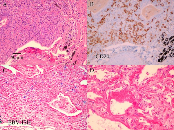
Figure 2.
A. Hematoxylin and eosin stain of the lung biopsy shows a polymorphic lymphoid infiltrate composed of large atypical cells, small lymphocytes and many plasma cells, with lymphoid cells infiltrating blood vessels and bronchial walls. (Scale bar in A also applies to B-D, original magnification 200×) B. Immunohistochemistry on a section adjacent to A shows that that many large atypical cells are positive for CD20 (B cell marker). C. In situ hybridization for Epstein-Barr virus (EBV) encoded RNA (EBER) on a section adjacent to A shows that many large lymphocytes are positive for EBER (stained blue). D. A chest-only autopsy revealed diffuse alveolar damage in both lungs, with areas of edema, fibrin deposition, and hyaline membrane formation. There is no evidence of residual lymphomatoid granulomatosis.

