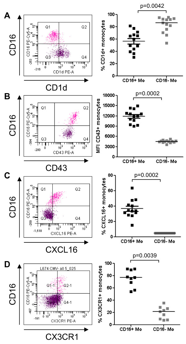
Figure 4.
Differential expression of CD1d, CD43, CXCL16, and CX3CR1 on CD16+ and CD16- monocytes. Freshly isolated PBMC were stained with Pacific Blue CD3, Alexa700 CD4, FITC CD14, PE-Cy5 CD16, and PE CD1d, PE CD43, PE CXCL16 or PE CX3CR1 Abs. Gated CD3-CD4lowCD14highCD16- (CD16- Mo) and CD3-CD4lowCD14lowCD16+ (CD16+ Mo) cells were analyzed for expression of (A) CD1d, (B) CD43, (C) CXCL16, and (D) CX3CR1. Shown are representative dot plots (left panels) and results for 9–13 different donors (right panels). Paired Wilcoxon signed rank test was used to calculated statistical significance (p < 0.05, CD16+ versus CD16- Mo).

