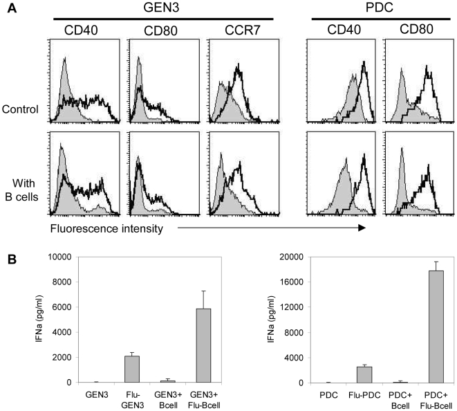
Figure 5. Activation of PDC upon exposure to flu-B cells.
(A) In upper panels, GEN3 or purified PDC were incubated (open curves) or not (filled curves) with influenza virus for 24 hours, as positive control of their maturation, and cells were stained with indicated mAb. In lower panels, B cells were exposed (open curves) or not (filled curves) to virus for 18 hours, washed, and then incubated with GEN3 or purified PDC. After a 24-hour co-culture, cells were stained with indicated mAb, and maturation was assessed by flow cytometry after gating on GEN3 cells (according to their FSC/SSC profile) or on BDCA2pos BDCA4 pos PDC. Data are representative of two independent experiments. (B) GEN3 or purified PDC were incubated with or without influenza virus, B cells or Flu-B cells for 24 hours, and then IFNα levels in supernatants were measured by ELISA. Data represent mean±SD of 2 experiments performed in duplicates.

