
Figure 2.
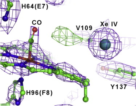
Xenon binding in the distal heme pocket of CO-Ngb electron density map in the XeIV niche, for the carbonmonoxy-Ngb•Xe structure (2Fo-Fc map, contoured at 1.7σ). XeIV is depicted as a sphere. The Fo-Fc map at 3.3σ is also displayed in green, showing an unmodeled positive electron density, which may be due to a small fraction of photodissociated CO (see Results).