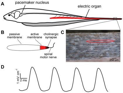
Figure 1. Generation of the EOD.
(A) The EOD is produced by the coordinated APs of the electric organ cells, called electrocytes. A medullary pacemaker nucleus controls the electrocyte APs via spinal electromotor neurons which innervate the electrocytes. (B) Electrocytes are innervated on the posterior end of the cell, where the spinal nerve forms a large cholinergic synapse. The electrically excitable region of the cell membrane, populated by Na+ and K+ channels, is localized to the posterior most region of the cell, extending approximately 150 µm toward the anterior of the cell. The remainder of the cell membrane is electrically passive. APs in the electrocytes cause current to move along the rostral-caudal body axis and out into the surrounding water. (C) A section of electric organ from the tail, with skin removed to expose the electrocytes, which are densely packed within the electric organ. A single electrocyte is outlined in red. (D) The EOD waveform recorded from S. macrurus is a sinusoidal wave emitted at a steady frequency by each fish. The EOD frequency among fish has a range of approximately 70 to 150 Hz.

