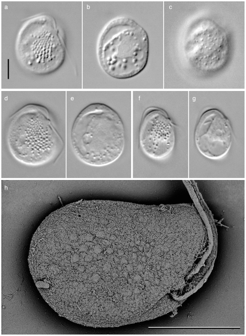
Figure 1. Light and scanning electron micrographs of Roombia truncata. sp. nov.
a–c. Holotype of R. truncata; d–e a cell showing size close to the maximum size; f–g. a cell showing size close to the minimum size. The ventral side (a, d, f) of the cell has 5–11 rows of conspicuous ejectisomes, whose diameter ranging from c.a. 0.3 µm at the anterior end and 0.7 µm at the posterior end. Smaller ejectisomes are also present on the dorsal face of the cell (c). A cell has the anterior and posterior flagella emerging from a papilla like structure of the ventral left subapical region (a, d, f), and food vacuole along the right margin of the cell (b, e, g). (h). scanning electron micrograph showing ventral side of the cell. Note multiple rows of ejectisomes. Scale bar = 5 µm.

