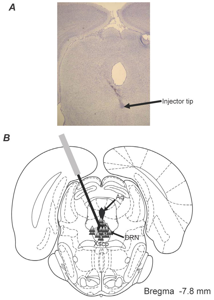
Figure 3.
(A) Histological example of an injector tip placed within the DRN. (B) Schematic representation of a coronal section of the rat brain depicting the distribution of DRN microinfusion sites. This section was taken from Paxinos & Watson (1998). Triangles denote placements within the DRN (shaded). Relevant anatomical structures are: Aq, aqueduct; Xscp, decussation of the superior cerebellar peduncle.

