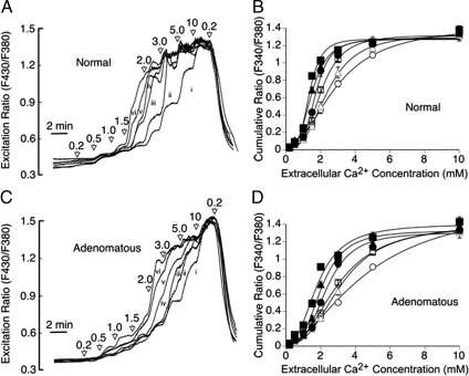
Figure 5.
Effect of l-Phe on Ca2+-dependent intracellular Ca2+ mobilization from normal and adenomatous parathyroid cells. The effect of stepwise increments in Ca2+o (from 0.2 to 10 mm) in the absence (i) or presence of 0.1 (ii), 0.3 (iii), 1.0 (iv), 3.0 (v) and 10 mm (vi) l-Phe are shown for fura-2 loaded normal (A) and adenomatous (C) parathyroid cells together with the impact of variations in l-Phe concentration on the Ca2+o concentration dependence of intracellular Ca2+ mobilization from normal (B) and adenomatous (D) parathyroid cells. The data have been corrected for baseline. The symbols are: ○, control; ▵, 0.1 mm l-Phe; □, 0.3 mm l-Phe; •, 1.0 mm l-Phe; ▴, 3.0 mm l-Phe; and ▪, 10 mm l-Phe. The results were obtained from experiments performed on six normal and eight adenomatous preparations, respectively. The traces in panels A and C represent means derived from 60 and 80 single cells per concentration of l-Phe tested, respectively.

