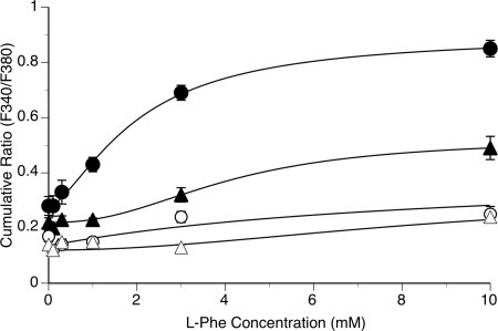
Figure 6.
Effects of l-Phe concentration on intracellular Ca2+ mobilization in normal and adenomatous parathyroid cells at Ca2+o concentrations of 1.0 and 1.5 mm. Normal and adenomatous parathyroid cells were prepared and loaded with fura-2 as described in Materials and Methods. The cells were then exposed to stepwise increments in Ca2+o concentration in the absence or presence of various concentrations of l-Phe (0.1–10 mm) as shown in Fig. 5. The symbols are: ○, Ca2+ 1.0 mm, normal; ▵, Ca2+ 1.0 mm, adenomatous; •, Ca2+ 1.5 mm, normal; and ▴, Ca2+ 1.5 mm, adenomatous. The data were obtained from cells prepared from parathyroid tissue samples from six patients who underwent thyroid surgery (normals) and eight patients with parathyroid adenomatous disease and primary hyperparathyroidism.

