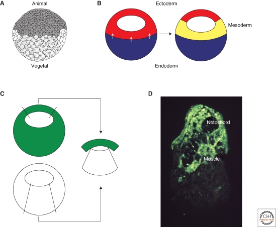
Figure 1.
Mesoderm induction. (A) Drawing of a Xenopus embryo at the midblastula stage (Nieuwkoop 1956). The animal hemisphere is to the top and the vegetal hemisphere to the bottom. The mesoderm forms in the equatorial region of the embryo between the two. (B) Mesoderm induction. A signal from the vegetal hemisphere of the embryo (white arrows) causes equatorial cells to form mesoderm. (C) Demonstration of mesoderm induction. Animal pole tissue derived from an embryo uniformly labeled with a fluorescent lineage label is juxtaposed with vegetal pole tissue from an unlabeled embryo and the resulting conjugate is cultured for 3 days. (D) The result of such an experiment. The fluorescently labeled tissue differentiates as notochord and muscle rather than epidermis. Photograph courtesy of Les Dale and Jonathon Slack.

