
Figure 1. Expression of Immature E-cadherin in A431DE Cells.
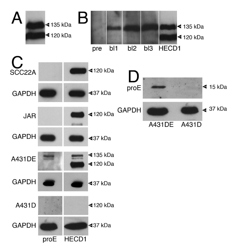
A. 50 µg of protein from TNE extracts of A431D cells transfected to overexpress E-cadherin (A431DEplasmid) was resolved by 7% SDS-PAGE and immunoblotted with the HECD1 monoclonal anti-E-cadherin antibody. The 135 kDa pro-region containing E-cadherin and the 120 kDa mature E-cadherin are pointed out. B. 50 µg of protein from TNE extracts of A431DEplasmid cells was resolved by 7% SDS-PAGE and immunoblotted with preimmune serum (pre) and successive bleeds (bl1, bl2, bl3) from a rabbit immunized with E-cadherin proregion peptide. Lane 5 shows an immunoblot of the same extract using HECD1 to point out mature (120 kDa) and immature (135 kDa) E-cadherin. C. 50 µg of protein from TNE extracts of SCC22A cells, JAR cells, A431DE cells (infected to express physiological levels of E-cadherin) and A431D cells were resolved by 7% SDS-PAGE and immunoblotted with anti-proE-cadherin antiserum or HECD1 monoclonal anti-E-cadherin antibody. GAPDH was used as a loading control. D. Lysates from A431DE and A431D cells were resolved by 15% SDS-PAGE and immunoblotted using anti-pro-E-cadherin antiserum. GAPDH was used as a loading control.