
Figure 3. Catenins Associate with Immature E-cadherin.
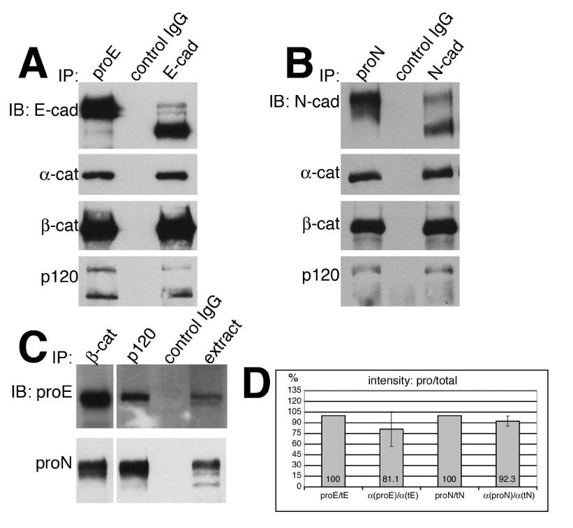
Various quantities of TNE extracts from A431DE (A) or A431DN (B) cells were immunoprecipitated with anti-pro-E-cadherin antiserum (800 µg protein from TNE extract), HECD-1 anti-E-cadherin monoclonal antibody (50 µg protein from TNE extract), 10A10 anti-pro-N-cadherin monoclonal antibody (700 µg protein from TNE extract), 13A9 anti-N-cadherin monoclonal antibody (30 µg protein from TNE extract) or control IgG (800 µg protein from A431DE TNE extracts or 700 µg protein from A431DN TNE extracts). The immunoprecipitation reactions were resolved by 7% SDS-PAGE and immunoblotted with HECD-1 anti-E-cadherin, 13A9 anti-N-cadherin, 1G5 anti-α-catenin, 15B8 anti-β-catenin, or pp120 anti-p120ctn. Both pro-E-cadherin and pro-N-cadherin were efficiently associated with αcatenin, β-catenin, and p120ctn. C. Extracts from A431DE cells (top panel) or A431DN cells (bottom panel) were immunoprecipitated using anti-β-catenin, or anti-p120ctn and immunoblotted with anti-pro-E-cadherin antiserum (top panel) or anti-pro-N-cadherin monoclonal antibody 10A10. D. TNE extracts from A431DE or A431DN cell lines were immunoprecipitated with anti-pro-E-cadherin, anti-E-cadherin, anti-pro-N-cadherin, or anti-N-cadherin as above and the immunoprecipitates were resolved by SDS-PAGE and immunoblotted with anti-E-cadherin, anti-N-cadherin, or anti-α-catenin. Bands were visualized by infrared fluorescent secondary antibodies using the Odyssey Imaging System to yield quantitative results. Shown is a bar graph with normalized ratios comparing intensity readings of α-catenin bound to pro-cadherin or total cadherin molecules. The data represents averages of three separate experiments.