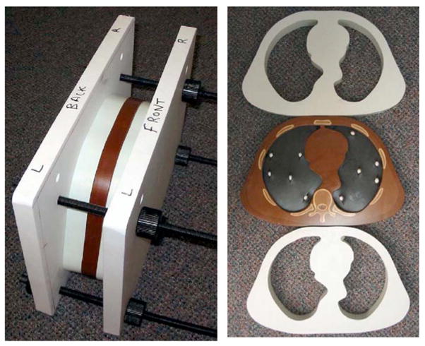
Fig. 1.

Left: Setup for CT scanning showing thorax section sandwiched between two water-equivalent bolus sections. The sections are tightly squeezed together using the outer plastic vice. Right: Front views of the two bolus sections and thorax section containing the 9.5 mm diameter spherical reference nodules with calcium carbonate concentrations of 50 mg/cc in the lung simulating foam on one side and 100 mg/cc on the other side.
